Novel X-ray technology improves contrast in soft tissue
Compact synchrotron makes tumors visible
2015-04-30 – News from Physics Department
X-ray images have become an integral part of daily medical practice. Bones, for example, absorb large amounts of X-rays because of their high calcium content. This allows them to be differentiated from air-filled cavities like the lungs and surrounding soft tissue. However, because of their very similar absorption coefficients, soft tissue, organs and structures inside organs, like tumors, are hardly discernable from one another using the medical devices deployed in medicine today.
Now a group of scientists headed by Franz Pfeiffer, Professor of Biomedical Physics in the Physics Department Department and the Faculty of Medicine at TU München, have for the first time succeeded in making such soft tissue visible. The scientists used a new kind of X-ray source that was developed only a few years ago.
Compact synchrotron source
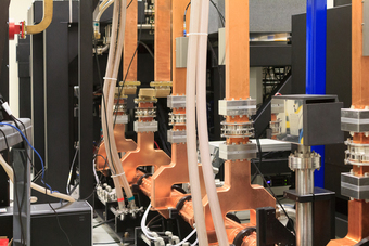
Unlike classical X-ray tubes, a synchrotron generates highly focused, monochromatic X-rays. The individual rays all have the same energy and wavelength. In the past, X-rays with these properties could only be generated in large particle accelerators, which have a circumference of at least one kilometer. The compact synchrotron, in contrast, has merely the size of a car and fits into a normal laboratory.
“Monochromatic radiation is much better suited for measuring other parameters, in addition to absorption,” explains Elena Eggl, doctoral candidate at the Chair of Biomedical Physics. “This is because it does not lead to artifacts that deteriorate the image quality.”
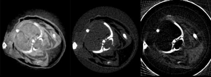
The scientists inserted an optical grating into the focused X-ray beam, allowing them to detect even tiniest phase shifts and scattering of the radiation in addition to the absorption of X-rays. The first phase contrast tomography image from a compact synchrotron source was successfully acquired.

Complementary information
The phase contrast, dark field and absorption images made using the new technology have complementary properties. Liquid in tissue that remains indiscernible and, thus, invisible using conventional X-ray tubes, suddenly comes to life. The greatly improved soft tissue contrast of the new X-ray technology could also help make tumors detectable earlier on and enable quick diagnoses – in medical emergencies, for example.
The clarity of the new technology becomes apparent when comparing white and brown fatty tissue. “In a mouse we were able to recognize not only heart, liver and other organs much better, but could even differentiate between brown and white body fat,” says Eggl.
Brown fatty tissue, which occurs mainly in newborns, can support the burning of normal white fatty tissue. It is a relatively new discovery that adults, too, still have brown fatty tissue. Tissue that – as some researchers hope – can be reactivated to help obese people lose weight.
While these experiments were performed using an initial prototype setup of Lyncean Technologies Inc. in California, a significantly improved compact synchrotron source is under construction at the Garching Research Campus. It is part of the “Center for Advanced Laser Applications” (CALA), a joint project of the TU München and the Ludwig-Maximillians Universität (LMU). Eggl and Pfeiffer, in collaboration with colleagues in laser physics at the LMU and the Max Planck Institute of Quantum Optics, hope to further improve the new X-ray technology.
The research was funded by the German Research Foundation via the Cluster of Excellence Munich-Centre for Advanced Photonics (MAP), the European Research Council (ERC, Starting Grant Nr. 240142), the National Institute of General Medical Sciences (USA, Grant R44-GM074437) and the National Center for Research Resources (USA, Grant R43-RR025730). Further cooperation partners included the Helmholz Center NanoMikro at the Karlsruhe Institute of Technology (KIT), the University of Lund (Sweden) and Lyncean Technologies Inc. (USA).
Publication
Contact
- Prof. Dr. Franz Pfeiffer
- Technische Universität MünchenJames-Franck-Str. 185748 GarchingTel.: +49 89 289-10807E-Mail: franz.pfeiffer@tum.de
