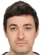
Dr. rer. nat. Florian Schaff
- Phone
- +49 89 289-10802
+49 89 289-12844 - Room
- –
- florian.schaff@tum.de
- Links
-
Page in TUMonline
- Group
- Biomedical Physics
Courses and Dates
Offered Bachelor’s or Master’s Theses Topics
- Convolutional neural networks and transfer learning for artefacts reduction in X-ray dark-field CT
Grating-based X-ray dark-field (DF) imaging uses scattering of X-rays to create an image of an object, rather than conventional X-ray attenuation. The combination of X-ray scattering with imaging allows us to map information about structures that are much smaller than the resolution of the imaging system over a large field of view. X-ray dark-field imaging can be combined with computed tomography (CT) to create three-dimensional images of the scattering distribution inside an object. DF-CT was recently implemented for the first time into a clinical CT here at TUM
(https://www.bioengineering.tum.de/en/news/details/new-technology-for-clinical-ct-scans).
The goal of this project is to use convolutional neural networks (CNNs) to remove sampling artefacts in DF-CT images. Due to the unavailability of training data from the DF-CT machine, a technologically similar experimental setup and apply transfer learning will be used. The student will acquire, process and prepare training data, as well as train and apply CNNs.
Character of thesis work: experimental lab work/ data acquisition (50%) & computational/ image processing (50%)
Basic experience in image processing, CNNs, and/or Python programming are desirable.
For more information, please contact: Dr. Florian Schaff (florian.schaff@tum.de), or Prof. Franz Pfeiffer (franz.pfeiffer@tum.de).
- suitable as
- Master’s Thesis Biomedical Engineering and Medical Physics
- Supervisor: Franz Pfeiffer
- Convolutional neural networks and transfer learning for artefacts reduction in X-ray dark-field CT
Grating-based X-ray dark-field (DF) imaging uses scattering of X-rays to create an image of an object, rather than conventional X-ray attenuation. The combination of X-ray scattering with imaging allows us to map information about structures that are much smaller than the resolution of the imaging system over a large field of view. X-ray dark-field imaging can be combined with computed tomography (CT) to create three-dimensional images of the scattering distribution inside an object. DF-CT was recently implemented for the first time into a clinical CT here at TUM
(https://www.bioengineering.tum.de/en/news/details/new-technology-for-clinical-ct-scans).
The goal of this project is to use convolutional neural networks (CNNs) to remove sampling artefacts in DF-CT images. Due to the unavailability of training data from the DF-CT machine, a technologically similar experimental setup and apply transfer learning will be used. The student will acquire, process and prepare training data, as well as train and apply CNNs.
Character of thesis work: experimental lab work/ data acquisition (50%) & computational/ image processing (50%)
Basic experience in image processing, CNNs, and/or Python programming are desirable.
For more information, please contact: Dr. Florian Schaff (florian.schaff@tum.de), or Prof. Franz Pfeiffer (franz.pfeiffer@tum.de).
- suitable as
- Master’s Thesis Applied and Engineering Physics
- Supervisor: Franz Pfeiffer
- Implementation of a grating-based interferometer for X-ray vector radiography at the Munich Compact Light Source
Grating-based X-ray dark-field (DF) imaging uses scattering of X-rays to create an image of an object, rather than conventional X-ray attenuation. The combination of X-ray scattering with imaging allows us to map information about structures that are much smaller than the resolution of the imaging system over a large field of view. The fact that the used gratings typically are one-dimensional can be leveraged to obtain an orientation dependent dark-field signal in a technique called X-ray vector radiography (XVR). Applications of XVR include the determination of the fibre orientation in reinforced composite materials, or characterization of the anisotropic structure in trabecular bones.
The goal of this project is to implement an experimental XVR setup at the Munich Compact Light Source (MuCLS - https://www.bioengineering.tum.de/en/central-building/munich-compact-light-source). The student will help with the design, implementation, and characterization of the X-ray grating interferometer setup, and conduct their own XVR experiments.
Character of thesis work: experimental lab work/ controls/ data acquisition (50%) & computational/ simulation/image processing (50%)
Basic experience in X-ray imaging, and/or Python programming are desirable.
For more information, please contact: Dr. Florian Schaff (florian.schaff@tum.de), or Prof. Franz Pfeiffer (franz.pfeiffer@tum.de).
- suitable as
- Master’s Thesis Biomedical Engineering and Medical Physics
- Supervisor: Franz Pfeiffer
- Implementation of a grating-based interferometer for X-ray vector radiography at the Munich Compact Light Source
Grating-based X-ray dark-field (DF) imaging uses scattering of X-rays to create an image of an object, rather than conventional X-ray attenuation. The combination of X-ray scattering with imaging allows us to map information about structures that are much smaller than the resolution of the imaging system over a large field of view. The fact that the used gratings typically are one-dimensional can be leveraged to obtain an orientation dependent dark-field signal in a technique called X-ray vector radiography (XVR). Applications of XVR include the determination of the fibre orientation in reinforced composite materials, or characterization of the anisotropic structure in trabecular bones.
The goal of this project is to implement an experimental XVR setup at the Munich Compact Light Source (MuCLS - https://www.bioengineering.tum.de/en/central-building/munich-compact-light-source). The student will help with the design, implementation, and characterization of the X-ray grating interferometer setup, and conduct their own XVR experiments.
Character of thesis work: experimental lab work/ controls/ data acquisition (50%) & computational/ simulation/image processing (50%)
Basic experience in X-ray imaging, and/or Python programming are desirable.
For more information, please contact: Dr. Florian Schaff (florian.schaff@tum.de), or Prof. Franz Pfeiffer (franz.pfeiffer@tum.de).
- suitable as
- Master’s Thesis Applied and Engineering Physics
- Supervisor: Franz Pfeiffer