New state-of-the-art compact X-ray source
World’s first mini particle accelerator for high-brilliance X-rays at TUM
2015-10-29 – News from the Physics Department

Scientists and physicians are still routinely using X-rays for diagnostic purposes 120 years after their discovery. A major aim has therefore been to improve the quality of X-rays in order to make diagnoses more accurate. For example, soft tissues could thereby be visualized better and even minute tumors detected. For a considerable time, a team at the Technical University of Munich (TUM) headed by Professor Franz Pfeiffer, Chair of Biomedical Physics, has been developing new X-ray techniques.
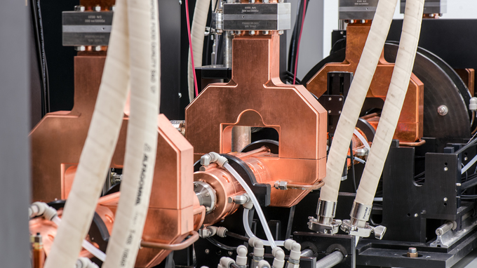
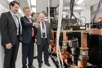
Starting October 29th, the scientists will now be able to use the world’s first mini synchrotron for high-brilliance X-rays at their institute. The Munich Compact Light Source (MuCLS) is part of the new Center for Advanced Laser Applications (CALA), a joint project between TUM and Ludwig-Maximilians-Universität München (LMU).
New technique: collision between electrons and a laser beam

The California-based company Lyncean Technologies, which developed the mini synchrotron, employed a special technique. Large ring accelerators generate X-rays by deflecting high-energy electrons with magnets. They obtain high energies by means of extreme acceleration, and this requires big ring systems.
The new synchrotron uses a technique where X-rays are generated when laser light collides with high-speed electrons – within a space that’s half as thin as a human hair. The major advantage of this approach is that the electrons can be traveling much more slowly. Consequently, they can be stored in a ring accelerator less than five meters in circumference, whereas synchrotrons need a circumference of nearly one thousand meters.
“We used to have to reserve time slots on the large synchrotrons long in advance if we wanted to run an experiment. Now we can work with a system in our own laboratory – which will speed up our research work considerably,” says Pfeiffer.
More intense, more variable and with better resolution
Apart from being more compact, the new system has other advantages over conventional X-ray tubes. The X-rays it produces are extremely bright and intense. Moreover, the energy of the X-rays can be precisely controlled so that they can be used, for example, for examining different tissue types. They also provide much better spatial resolution, as the source of the beam is less diffuse thanks to the small collision space.
“Brilliant X-rays can distinguish materials better, meaning that we will be able to detect much smaller tumors in tissue in the future. However, our research activities also include measuring bone properties in osteoporosis and determining altered sizes of pulmonary alveoli in diverse lung diseases,” explains Dr. Klaus Achterhold from the MuCLS team.
The scientists will initially use the instrument mainly for preclinical research, i.e. examining tissue samples from patients. They will also combine the new X-ray source with other systems, such as phase contrast. The group headed by Franz Pfeiffer has developed and refined the new X-ray phase-contrast technique.
- Editing
- Dr. Vera Siegler, Dr. Johannes Wiedersich
Contact
- Dr. Klaus Achterhold
- Technische Universität MünchenJames-Franck-Str. 185748 GarchingTel.: +49 89 289-12559E-Mail: klaus.achterhold@ph.tum.de